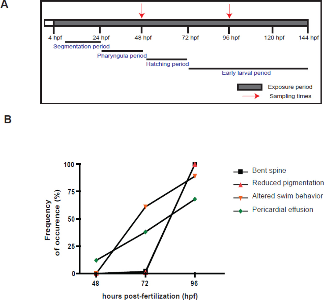
Figure 1.
(A). Outline of the experimental design. Zebrafish embryos were exposed to VPA (1 mM) and ethanol (solvent carrier control; 0.001% v/v) starting from 4 hours post-fertilization (hpf) continuously until the end of the experiment (144 hpf). Embryos were observed daily for developmental abnormalities and sampled at 48 and 96 hpf for microarray analysis. The exposure period is shown in shaded colors and the red arrows represent the two sampling time points. The key stages of zebrafish development are highlighted (Westerfield, 2000). (B). VPA-induced phenotypic defects during the course of exposure. Embryos were observed under the microscope for any phenotypic abnormalities every 24 hours until the end of the experiment. The different phenotypic defects observed include bent spine, reduced pigmentation, altered swimming behavior and pericardial effusion. The percentage of embryos with each defect was calculated by dividing the total number of embryos with the deformity by the total number in each replicate.

