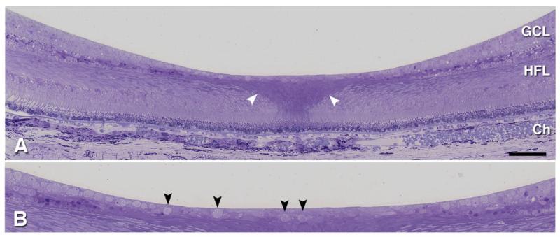
Figure 2.
Histology of the normal human fovea. 1 μm thick epoxy section through retina and choroid of 81 yr old woman, stained with toluidine blue: GCL, ganglion cell layer; HFL, Henle fiber layer; Ch, Choroid. A. Note the darker staining of the central bouquet of cone photoreceptors and Müller cells, sclerad to the foveal floor (white arrowheads). B. Excerpt from panel A, magnified view shows isolated ganglion cells within the foveal floor (black arrowheads). The “ganglion cell layer” consists of ganglion cells, displaced amacrine cells, and intercalated Müller cells.

