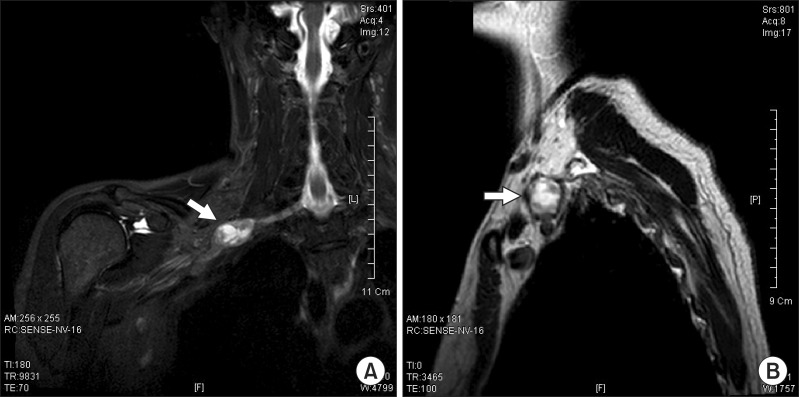
Fig. 2.
(A) T2 coronal magnetic resonance imaging (MRI) and (B) T2 sagittal MRI right brachial T2 coronal view images indicates an approximately 3.0×1.8×1.7 cm ovoid mass (arrow) between the inferior trunk and the anterior division of the brachial plexus (A). At the lateral arc of the right first rib, the T2 sagittal view also revealed an ovoid mass (arrow).

