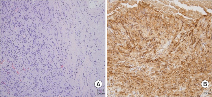
Fig. 3.

(A) Hematoxylin and eosin-stained tumor composed of spindle-shaped neoplastic Schwann cells with alternating areas of compact, elongated cells with occasional nuclear palisading (Antoni A region on the left field) and less cellular, loosely textured areas (Antoni B region on the right field) (×100). (B) Immunohistochemical staining (×300 magnification) demonstrated that the tumor cells were positive for S-100 protein.
