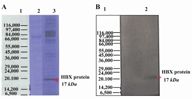
Figure 2.
Expression of HBX in Hep G2 cell line by SDS PAGE and Western blot. The purified bands had a molecular size of ~17 kDa, corresponding with the size of HBX protein; A) Hepatitis B virus X protein was separated on 12% SDS-polyacrylamide gel electrophoresis; Lane 1: Protein marker (Sigma marker, M 3913), Lane 2: Non-transfected cell (negative control), Lane 3: transfected cell; B) Expression of HBX 16.7 kDa was analyzed using Western blot by serum samples from patients with Liver Cirrhosis. Lane 1: Protein molecular weight marker (Sigma marker, M 3913), Lane 2: transfected cell with HBX

