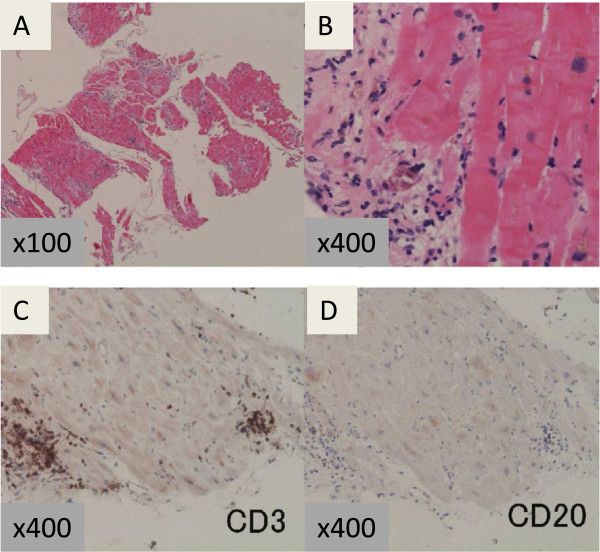
Figure 4.
Histopathological examination of a biopsy specimen from the left ventricle. Low-power view (× 100) (A) and high-power view (× 400) (B) of the biopsy specimen obtained from the left ventricle, stained with hematoxylin and eosin, showing lymphocytic infiltration into the interstitium and mild necrosis of the myocardium. Immunohistochemical staining with anti-CD3 (C) and anti-CD20 antibodies (D). CD3-positive lymphocytes were predominant.

