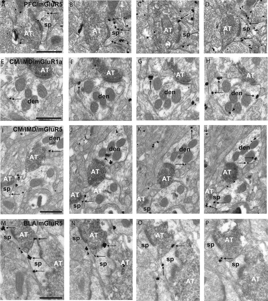
Figure 4.
Serial sections of rat accumbens tissue double immunostained for BDA (immunoperoxidase) and group I mGluRs (immunogold). A-D: Series of micrographs of a positively labeled axon terminal (AT) from the PFC forming an asymmetric synapse with a mGluR5-labeled spine (sp) in the accumbens core. Note that the majority of plasma membrane-bound labeling is extrasynaptic (single arrows), except for A, which shows an example of perisynaptic mGluR5 labeling (double arrowhead). E-H: Four micrographs of extrasynaptic mGluR1a labeling on a dendrite (den) contacted by an AT from theCM/IMD. I-L: Example of extrasynaptic mGluR5-IR in a dendrite and spine contacted by a positively labeled AT from the CM/IMD. There is also intracellular labeling in the dendrite (arrowheads). M-P: Example of mGluR5-IR spines in the accumbens shell contacted by labeled ATs from the posterior BLA with both extrasynaptic plasma membrane-bound labeling and intracellular labeling. Scale bar = 0.2 µm in A (applies to A-D), E (applies to E-H), I (applies to I-L), and M (applies to M-P).

