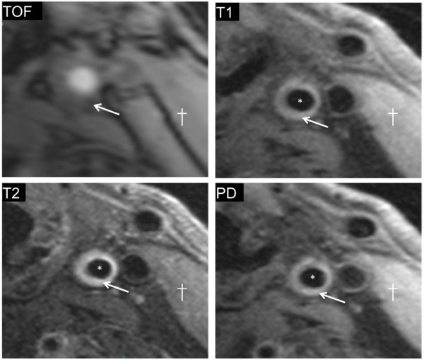
Figure 3.

Example of restenosis due to possible fibro-atheromatous narrowing. 50% restenosis of the left internal carotid artery imaged 4 years after surgery is shown. The signal appears hypointense on TOF images (arrow), but isointense signal is seen from the tissue surrounding the lumen (*) on the T1W image (arrow); the signal is similar in intensity to that from the adjacent sternocleidomastoid muscle. Similarly, on PDW images (arrow) the signal appears to be isointense compared to sternocleidomastoid (†), suggestive of fibrous tissue deposition.
