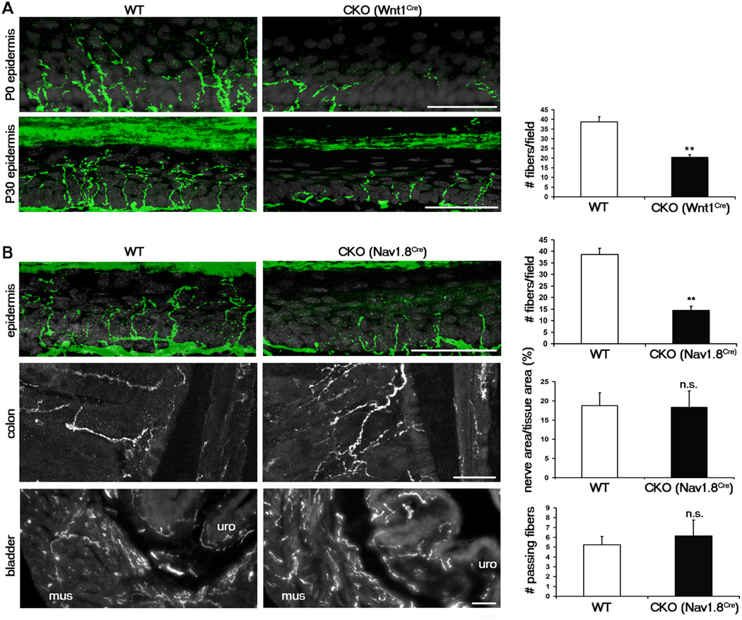
Figure 2. Runx1 is required selectively for sensory innervation to the epidermis.
(A) Sensory innervation to the thin glabrous epidermis of the hindpaw at P0 and P30 in the WT (Runx1F/F) and early CKO (Runx1F/F;Wnt1Cre), represented by PGP9.5 staining (green). Keratinocytes are represented by DAPI staining (grey). The number of PGP9.5+ fibers/field of 100 DAPI+ basal keratinocytes at P30 was quantitated.
(B) Sensory innervation to indicated regions of WT and late CKO mice at P30, by PGP9.5 staining for epidermis and by GFP immunostaining for colon and bladder. For the epidermis, “WT” is Runx1F/F, and “CKO” is Runx1F/F;Nav1.8Cre. The number of PGP9.5+ fibers/field of 100 DAPI+ basal keratinocytes at P30 was quantitated. For the colon and the bladder, “WT” is Nav1.8Cre;TauLSL-,EGFP/+, and “CKO” is Runx1F/F;Nav1.8Cre;TauLSL-,EGFP/+. The percentages of colon tissue area that is innervated and the number of fibers passing through lines drawn through the muscular layer of the bladder were quantitated. Two methods were used to quantitate bladder innervation, both producing similar results (see Supplementary Methods and Figure S5). n = 3 animals for each genotype. Error bars, SEM; ** p < 0.01; n.s. (not significant) p ≥ 0.05. Methods of quantitation are detailed in Supplementary Experimental Procedures and in Figure S5.
All scale bars represent 50 µm.

