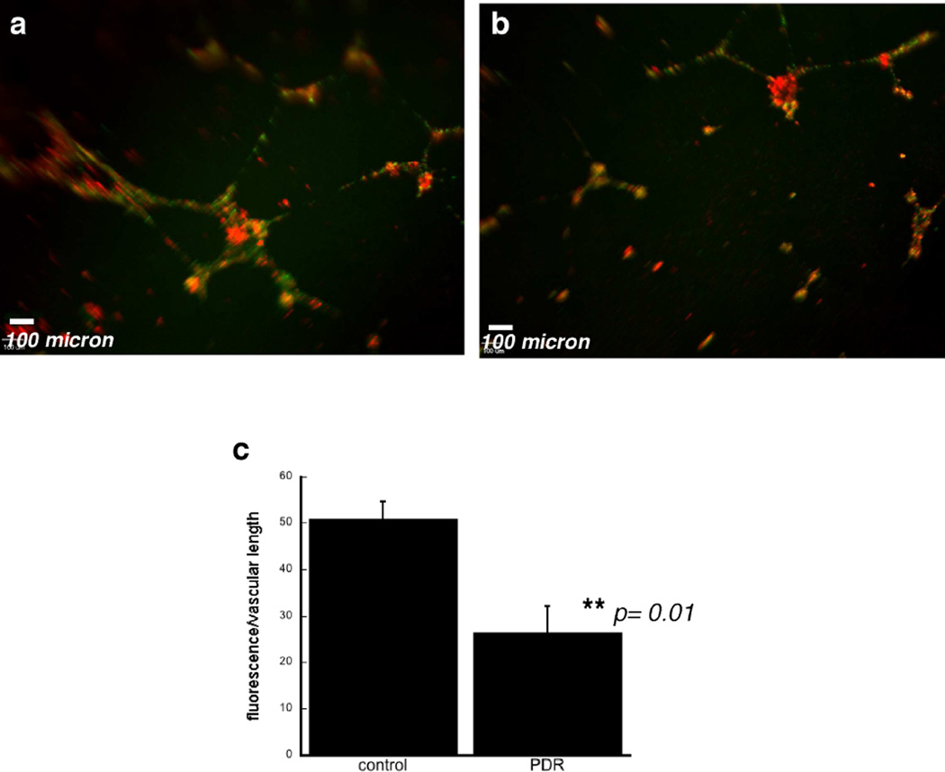
Figure 4. Co-culture of human retinal endothelial cells and ECFCs in an in vitro tube-forming assay.
Representative photomicrographs of human retinal endothelial cells labeled with Qdot®-525 (green) nm cell-tracking nanocrystals and co-cultured with ECFCs labeled with Qdot®-655 (red) nm cell-tracking nanocrystals from healthy controls (a) and patient with PDR (b). Scale bar (white) = 100 µm. A total of three PDR patients’ and three healthy control patients’ ECFCs were analyzed. c) quantitation of % ECFC fluorescence/vascular length was compared using ECFCs from patients with and without PDR.

