
Figure 5.
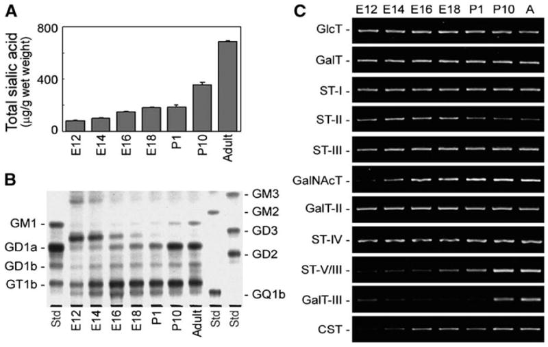
(A) Expression levels of gangliosides in the developing mouse brain. (B) Expression patterns of gangliosides in developing mouse brain analyzed by thin-layer chromatography. During development, the ganglioside expression patterns in mouse brain shifted from simple gangliosides such as GM3 and GD3 to complex gangliosides such as GM1 and GD1a. (C) Expression levels of GT genes in the developing mouse brain analyzed by RT-PCR. During development, GalNAcT (GA2/GM2/GD2/GT2-synthase) were increased, and ST-II (GD3-synthase) slightly decreased. “A” indicates adult mouse brain. (Reproduced from [66])