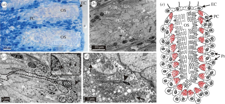
Figure 2.
Morphology of the starfish eye. (a) LM of two ommatidia sectioned longitudinally. Each of the fully developed ommatidia is composed of 100–150 photoreceptors and about the same number of pigment cells (PC). Note the thin layer of epithelial cells (EC, arrowhead) covering the outer segments (OS). The receptors are arranged in seven to eight layers perpendicular to the surface (b). (c) Longitudinal section of a photoreceptor (Pr, dashed black line). Interestingly, it receives feedback from the nervous system as indicated by afferent synapses (insert, arrowhead indicates synapse, arrows indicate synaptic vesicles). PrN, photoreceptor nucleus. (d) The outer segments of the starfish photoreceptors are morphologically a mixture of the rhabdomeric- and ciliary-type receptors. They are formed by microvilli coming both directly from the cell membrane (arrows) and from a modified cilium (arrowhead). (e) Schematic drawing of an ommatidium showing the layered arrangement of the photoreceptors and pigment cells.

