
Fig. 1. Ahr expression is silent in pluripotent mouse ES cells and activated during non-directed differentiation.
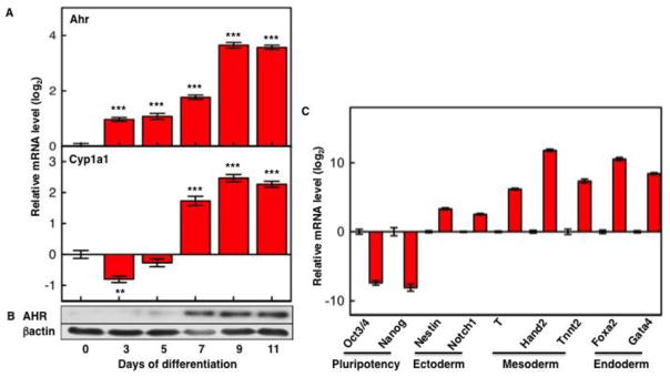
After 3-days of EB formation, differentiation was followed for 11 or 13 days on cover slips. Ahr and Cyp1a1 mRNA levels (A) were normalized to Gapdh and are shown relative to the levels in ES cells (day 0). Immunoblots with AHR and β–actin specific primary antibodies (B) were visualized with chemiluminescence ECL Western Blot Substrate. (C) mRNA expression levels of pluripotency and germ cell layer marker genes were examined by real time PCR, normalized to Gapdh and are shown relative to the levels in ES cells. The asterisk (*) indicates significant differences to values in ES cells: (*) p<0.05; (**) p<0.01; (***) p<0.001.