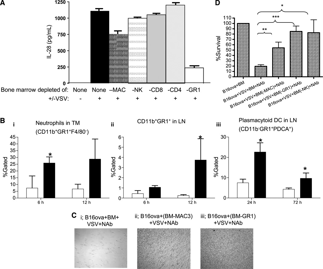
Figure 4.
GR1+ cells in bone marrow sense VSV. A, bone marrow from C57Bl/6 mice was left undepleted (none) or depleted of macrophages, NK, CD8, CD4, or GR1+ cells and cocultured with B16ova cells +/− VSV (MOI 0.1) as in Fig. 1A. Forty-eight hours after addition of VSV, supernatants were assayed for IL-28. B, C57Bl/6 (3 mice/group) bearing B16ova tumors were injected intratumorally with 5 × 10⁸ pfu of VSV (filled column) or PBS (opened column). Tumors (i) and draining lymph node (LN; ii and iii) were harvested at times shown and analyzed for (i) neutrophils (CD11b+GR1+F4/80−), CD11b+GR1+ (ii), or plasmacytoid dendritic cells (CD11b+GR1+PDCA+; iii). Infiltrates in tumors injected intratumorally with heat-inactivated VSV as a negative control were directly comparable with those tumors treated with PBS (data not shown). TM, tumor; DC, dendritic cells. C, C57Bl/6 bone marrow was left undepleted (i) or depleted of macrophages (ii) or GR1+ cells (iii), and cocultured with B16ova with VSV (MOI, 0.1) and neutralizing anti-VSV antibody (NAb) as in Fig. 1A. Forty-eight hours after the addition of VSV, cell survival was visualized or quantified by MTT assay (D). *, P < 0.05; **, P < 0.01; ***, P < 0.001.

