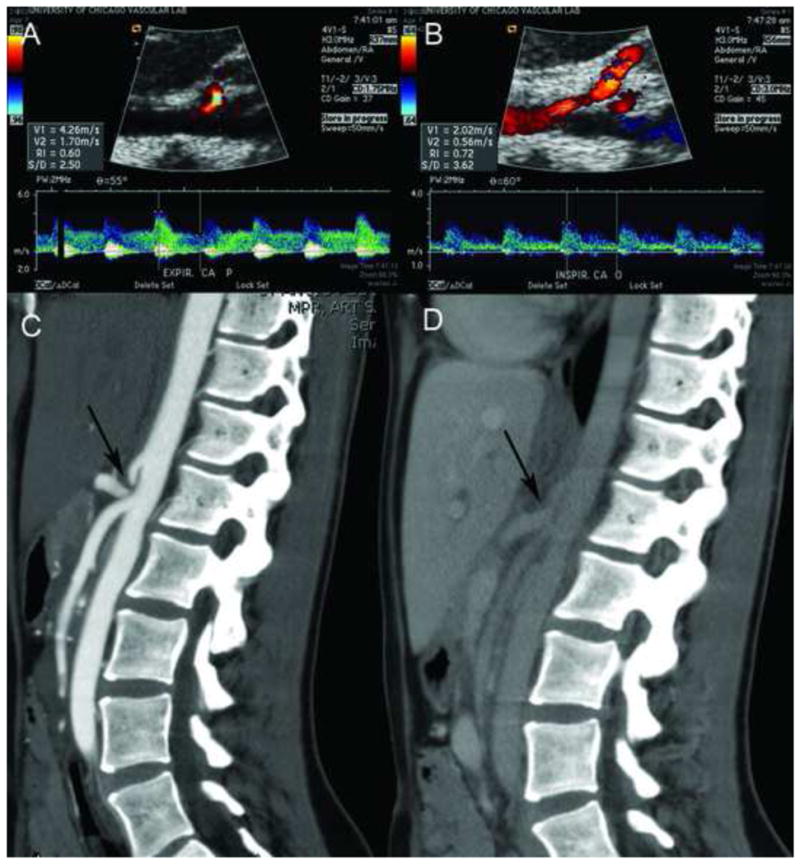
Figure 1.

Ultrasound with B-mode imaging showing the Aorta in logitudinal-section and arrow pointing at the origin of the celiac artery during expiration (A) with corresponding increase in peak systolic velocity to 426 cm/s. Figure 1B: The Aorta in logitudinal-section with the origin of the celiac artery during inspiration with a corresponding decrease in peak systolic velocity to 202 cm/s. Figure 1C: CT-Angiogram in sagittal view showing the origin of the celiac artery arising from the Aorta with a “J-Hook” configuration (arrow) during expiration and (D) during deep inspiration. Note the increase of the lumen size with deep inspiration (arrow) in D. The origin of the superior mesenteric artery is normal in appearance.
