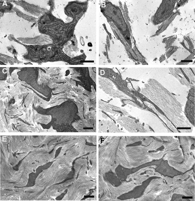
Fig. 1.
Transmission electron microscopy from ultrathin sections of Durcupan-embedded chick corneal stroma at embryonic days 10 (A and B), 14 (C and D), and 18 (E and F). Keratocytes adopt flattened morphology with extended cell processes. Collagen bundles appear at cell surfaces in cell recesses and parallel to cell processes at days 10 and 14. Bundles form a lamellar structure at day 18. (Scale bars, 2 µm, 500 nm in D.)

