
Fig. 7.
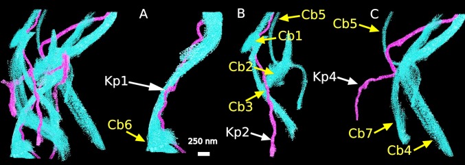
Surface rendering of a 3D reconstruction from day 12 chick cornea in meridional section reveals associations of collagen bundles (Cb) with individual keratocyte processes (Kp). (A–C) Structures isolated from the composite reconstruction (far left) demonstrate that some processes associate with only one collagen bundle (A, Kp 1 with Cb 6); others have multiple associations (B, Kp 2 with Cb 1, 2, 3, and 5; C, Kp 4 with Cb 4, 5, and 7). (Scale bar, 250 nm.)