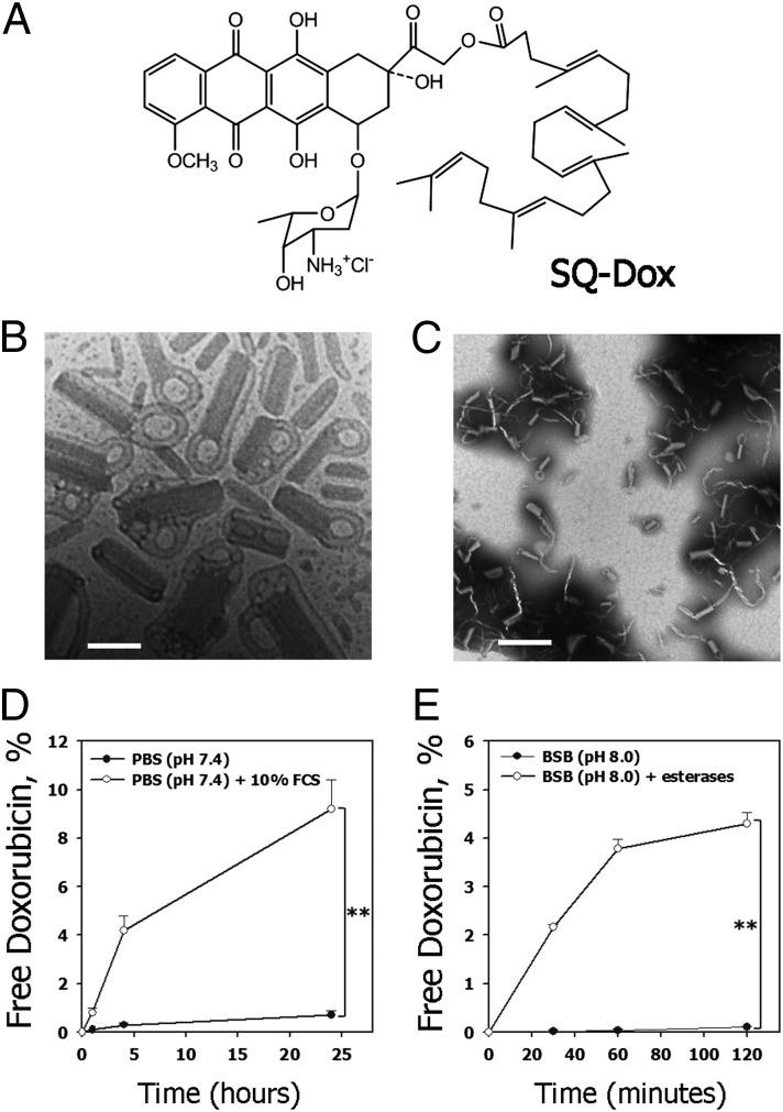
Fig. 1.
Nanoassemblies of the squalenoyl prodrug of doxorubicin (SQ-Dox NAs). (A) Chemical structure of SQ-Dox. (B) Cryo-TEM appearance of the SQ-Dox NAs. (Scale bar, 100 nm.) (C) TEM appearance of the SQ-Dox NAs. (Scale bar, 500 nm.) (D) Time course of doxorubicin release at 37 °C in PBS (pH 7.4) solution containing 10% FCS (n = 3, **P < 0.01). (E) Time course of doxorubicin release at 37 °C in borate (BSB, pH 8.0) solution containing 25 U/mL esterases (n = 3, **P < 0.01). Released doxorubicin was separated from the SQ-Dox NAs by centrifugation and quantified using HPLC (for experimental details, see SI Materials and Methods).

