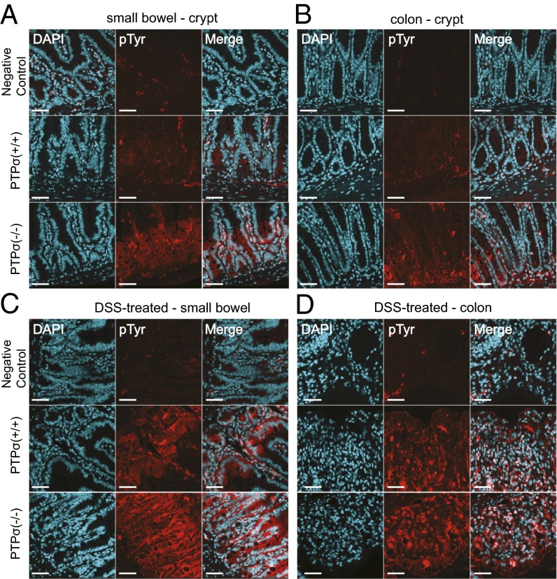
Fig. 3.
Tyr phosphorylation is enriched in the crypt regions of PTPσ−/− mouse colon and small bowel. Tissue sections from both naïve and DSS-treated mice were immunostained with anti-pTyr antibody and visualized using confocal microscopy. (A) Tyr phosphorylation is enriched in the crypt region of PTPσ−/− mouse small bowel compared with littermate controls. Increased Tyr phosphorylation is also observed in cells contained within the lamina propria regions of PTPσ−/− mice. (B) Increased Tyr phosphorylation is visible in the crypt regions of the colon in PTPσ−/− mice compared with littermate controls. (C and D) DSS treatment of the PTPσ−/− mice and littermates leads to increased Tyr phosphorylation in both KO and WT small bowel (C) and colonic (D) tissues. (Scale bars: 25 µm.)

