Fig. 6.
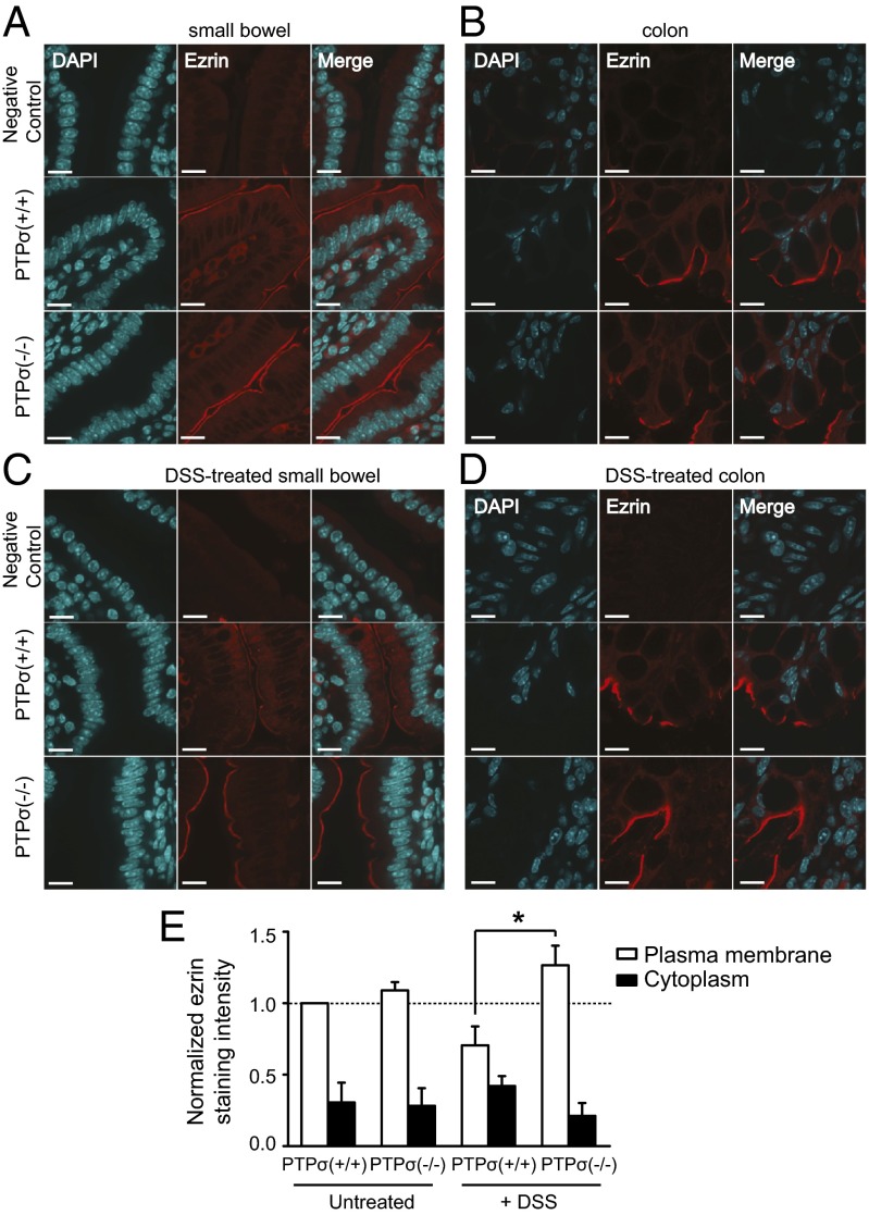
Ezrin localization is altered in the small bowel of PTPσ−/− mice after colitis-inducing DSS treatment. Ezrin localization was evaluated in small bowel and colon tissue sections from both naïve and DSS-treated PTPσ−/− mice and littermate controls using immunofluorescence microscopy. (A) Ezrin present at the apical PM of enterocytes in the small bowel in PTPσ−/− mice and controls. (B) Ezrin localizes to the apical surface of enterocytes in the colon, but there was no observed difference between PTPσ−/− mice and littermate controls. (C) Ezrin localization is disrupted in the small bowel of PTPσ−/− mice treated with DSS. (D) DSS treatment has no effect on the localization of ezrin in the colon. (Scale bars: 25 µm.) (E) Quantification of ezrin-staining intensity in enterocytes of both naïve and DSS treated PTPσ+/+ and PTPσ−/− mice. All staining-intensity values were normalized to the mean PM intensity observed in the naïve PTPσ+/+ mice. Images were converted to grayscale, and edges corresponding to the PM were isolated algorithmically. Data are means ± SD (n = 40–50 images per genotype; *P = 0.0046; Student t test).

