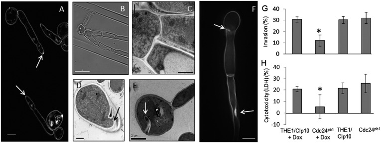
Fig. 4.
Growth and tissue penetration defects of Cdc24pb1 cells. (A) Hyphae stained with FM4-64 to show the Spitzenkörper (arrows). (Scale bars, 5 µm.) (B and C) Bifurcation of hyphae. (Scale bars, 10 µm and 1 µm.) (D and E) Mislocalized cell wall material in yeast and pseudohyphal cells (arrows). (Scale bars, 1 µm and 20 µm.) (F) Hypha stained with Calcofluor White with deposits of cell-wall material (arrows). (Scale bar, 5 µm.) (G) Yeast cells (1 × 10⁵) were inoculated onto confluent monolayers of H4 intestinal epithelial cells and incubated with or without 0.125 µg/mL doxycycline. Using a fluorescent anti-Candida antibody, hyphal cells were categorized as penetrating (no fluorescence) or nonpenetrating (fluorescent staining). (H) Cdc24pb1 and control strain yeast cells (2 × 10⁵ cells) were coincubated for 8 h with H4 cells with or without 0.125 µg/mL doxycycline and supernatants analyzed for lactate dehydrogenase release. *P ≤ 0.01. Error bars indicate SD.

