Fig. 3.
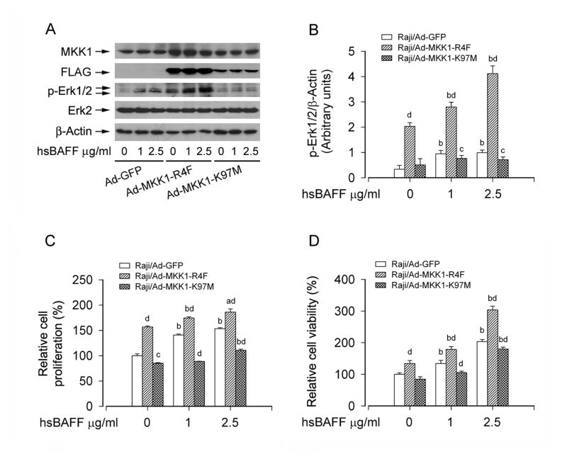
Expression of constitutively active or dominant negative MKK1 influences Erk1/2 activity and proliferation/viability in B cells. Raji cells, infected with Ad-MKK1-R4F, Ad-MKK1-K97M, and Ad-GFP (as control), respectively, were treated with 0, 1, 2.5 μg/mL hsBAFF for 12 h (for Western blotting) or 48 h (for cell proliferation/viability assay). Total cell lysates were subjected to Western blotting using indicated antibodies (A). The blots were probed for β-actin as a loading control. Similar results were observed in at least three independent experiments. The cell proliferation was evaluated by cell counting (C) and the cell viability was determined by the MTS assay (D). (A) Infection of Raji cells with Ad-MKK1-R4F and Ad-MKK1-K97M, but not Ad-GFP, resulted in expression of high levels of FLAG-tagged MKK1 mutants. Expression of MKK1-R4F resulted in robust phosphorylation of Erk1/2 even without stimulation with hsBAFF, whereas expression of MKK1-K97M suppressed hsBAFF-stimulated phosphorylation of Erk1/2. (B) Blots for p-Erk1/2 were semi-quantified using NIH image J. (C and D) Expression of MKK1-R4F significantly elevated the basal or hsBAFF-stimulated cell proliferation and viability, but expression of MKK1-K97M markedly inhibited these events. Results are presented as mean ± SE (n = 3–6). aP <0.05, bP <0.01, difference vs 0 μg/mL hsBAFF group; cP <0.05, dP <0.01, Ad-MKK1-R4F group or Ad-MKK1-K97M group vs Ad-GFP group.

