
Figure 2. Detection of IL-15 protein from granulocytic myeloid cells.
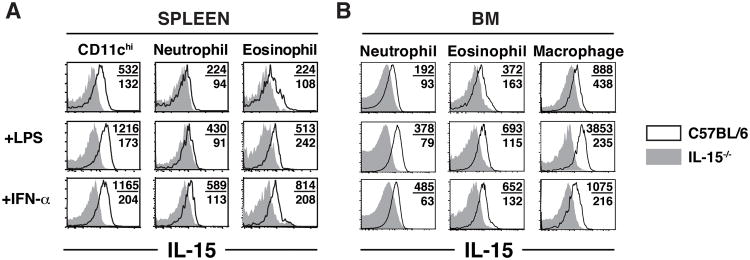
(A,B) 107 cells from the spleen (A) or BM (B) of WT (black lines) and IL-15-/- mice (gray filled histograms) were incubated O/N in the absence or presence of 1ug/ml LPS or 300U/ml IFN-α. Numbers indicate the MFI of IL-15 staining for WT (top) and IL-15-/- samples (bottom). Plots are representative of 2-3 experiments.