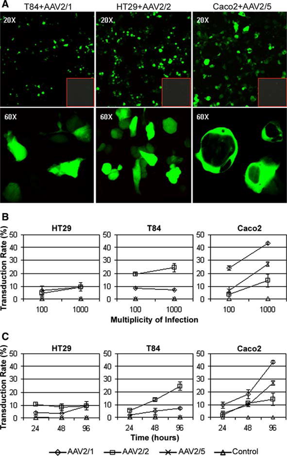
Fig. 1a–c.

Adeno-associated virus (AAV)-GFP transduced human colonic epithelial cell lines. a Representative confocal laser scanning microscopy images of live cells in culture expressing cytoplasmic GFP, 96 h after transduction with AAV-GFP at a particle multiplicity of infection (MOI) of 1,000, shown at 20× and 60× magnification. Red outlined insets Simultaneous negative control transductions. b, c Variation of dose curve and time course between cell lines. Cells were transduced with a particle MOI of 100 or 1,000 of AAV2/1, 2/2 & 2/5 encoding GFP. The percentage of GFP-positive cells (transduction rate) was quantified 96 h after transduction using flow cytometry (b). The transduction rate of cells transduced with MOI 1,000 was determined at 24, 48 and 96 h with each pseudotype (c). Data are presented as mean +/– SD of three separate experiments
