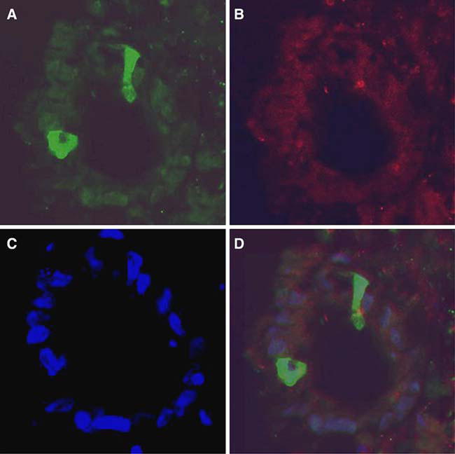
Fig. 3a–d.

Transduction of human colon biopsy tissue in organ culture with AAV2/2 encoding GFP and wt AdV coinfection. After frozen sectioning, GFP-positive epithelial cells are seen on cross-section of a villous at 40× magnification (a). The same image is seen under a rhodamine filter (b) for auto-fluorescence and a DAPI filter (c) for nuclear staining. Nonspecific or yellow staining was not seen in the overlay image (d)
