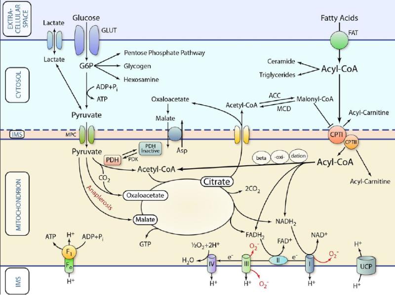
Figure 1. Schematic representation of classic pathways of cardiac metabolism.
Substrates are transported across the extracellular membrane into the cytosol and are metabolized in various ways. For oxidation, the respective metabolic intermediates (e.g., pyruvate or acyl-CoA) are transported across the inner mitochondrial membrane by specific transport systems. Once inside the mitochondrion, substrates are oxidized or carboxylated (anaplerosis) and fed into the Krebs cycle for the generation of reducing equivalents (NADH2 and FADH) and GTP. The reducing equivalents are used by the electron transport chain to generate a proton gradient, which in turn is used for the production of ATP. This principal functionality can be affected in various ways during HF thereby limiting ATP production or affecting cellular function in other ways (see text and further Figures for details). IMS: mitochondrial intermembrane space; GLUT: glucose transporter; FAT: fatty acid transporter. MPC: mitochondrial pyruvate transporter. (Illustration Credit: Ben Smith)

