
Figure 1. TAL effector structure.
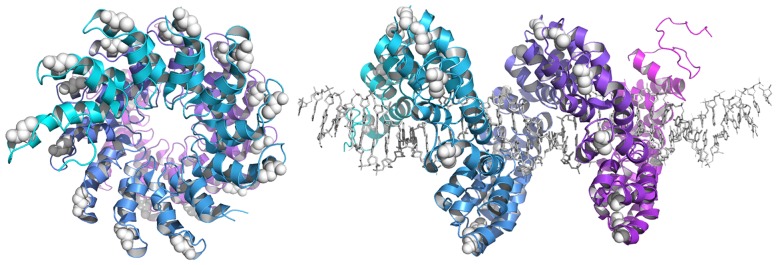
(Left) Front view of the PthXo1 DNA-binding domain in the absence of target DNA and (right) side view in the presence of target DNA. Surface-exposed Cys residues depicted as white spheres. TAL effector repeats are colored cyan and purple. DNA is shown as grey sticks. PDB ID: 3UGM [41].