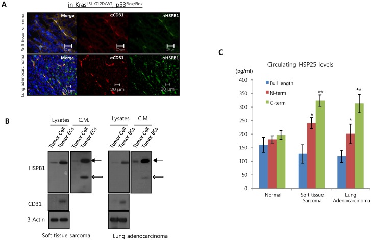
Figure 1. Soluble HSPB1 is secreted by tumor endothelial cells.
A Co-localization of HSBP1 and CD31 on tumor vessels in soft tissue sarcomas and lung adenocarcinomas derived from KrasLSL-G12D/WT; p53Flox/Flox mice was detected by immunofluorescence. B Tumor cells and tumor endothelial cells (ECs) were isolated from sarcomas and lung adenocarcinomas of KrasLSL-G12D/WT; p53Flox/Flox mice and cell lysates were analyzed by western blot. Secreted HSPB1 was detected in conditioned media (C.M.) from these cells. arrows, full length HSPB1; open arrows, cleaved HSPB1 C Serum HSPB1 levels in KrasLSL-G12D/WT; p53Flox/Flox mice with or without tumors were measured by ELISA assays for detecting intact HSPB1 as described in Methods; blue bars, anti-HSPB1 (10-21)+anti-HSPB1 (158-205) antibody. Red and green bars obtained from general ELISA assays using antibodies to the N- (10-21) or C-termini (158-201) of HSPB1 (*P<0.05 and **P<0.01 vs intact HSPB1).

