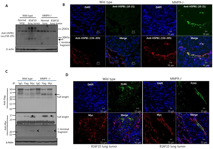
Figure 6. HSPB1 cleavage by MMP9 occurs during tumor progression.
A B16F10 melanoma cell lines were injected into the tail vein of wild-type and MMP9-null mice. 14 days later, lung tumor tissues were harvested. HSPB1 levels in the lung tissue samples were analyzed by western blotting using the HSPB1 (158–205) antibody. B Immunofluorescence staining of the N-terminal and C-terminal HSPB1 fragments in normal and B6F10 lung tissues using the HSPB1 (10–21) and (158–205) antibodies, respectively. C Lung tumor tissues were obtained via intravenous injection of B6F10 cells secreting wild-type HSPB1 into wild-type or MMP9-null mice. Immunoprecipitates with control IgG, anti-Flag, and anti-Myc in lysates of lung tumor tissues were subjected to western blot analysis using the indicated antibodies. D Immunofluorescence staining of the N-terminal and C-terminal HSPB1 fragments in B6F10 lung tumor tissues, using the Flag and Myc antibodies, respectively.

