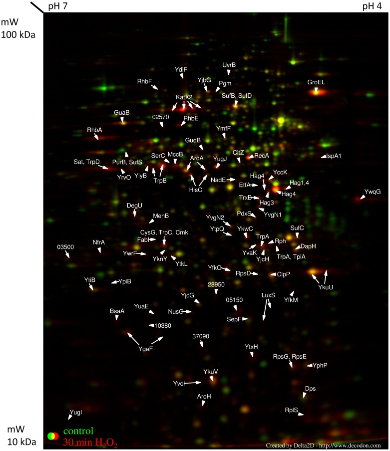
Figure 4. Cytosolic proteome 30 min after H2O2 treatment.
The cytosolic proteome of B. pumilus cells 30 min after H2O2 treatment. Cell samples were labeled with L–[35S]-methionine during the exponential growth phase (OD500 nm 0.6), and 30 min after H2O2 addition. Proteins were separated in a pH gradient 4 (right) –7 (left).

