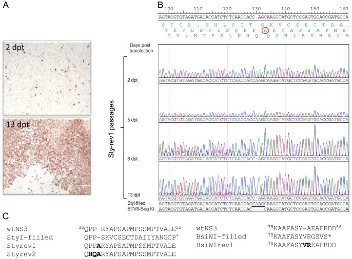
Figure 2. Analysis of revertant viruses Sty-rev and BsiWI-rev.
(A) anti-VP7 immunostained cells after 2 or 13 days post transfection with BTV1 Seg-1-9 and mutated Seg-10 StyI-filled resulting in revertant virus StyI-rev1 showing CPE at 13 dpt. (B) sequence analysis of Seg-10 amplicons from StyI-rev1 passages at different time points after transfection. In single letter code, putative translation of all three frames is shown of which the middle is the ORF of NS3 with the insertion of Alanine (A) in StyI-rev1. Here the analysis is shown by use of a sequence primer located downstream of the StyI site. Note the mixed sequence at 8 dpt upstream of the filled site due the introduction of the point deletion at the StyI site in a subpopulation of the fragments. (C) Comparison of the amino acid sequences of NS3 of wtBTV1/8(S10), the 4-basepairs insertions, and the rescued revertant viruses Sty-rev1, 2 and BsiWI-rev1. Amino acids changes in the regions of the 4-basepairs insertion of the revertant viruses Sty-rev1, 2 and BsiWI-rev1 are in bold.

