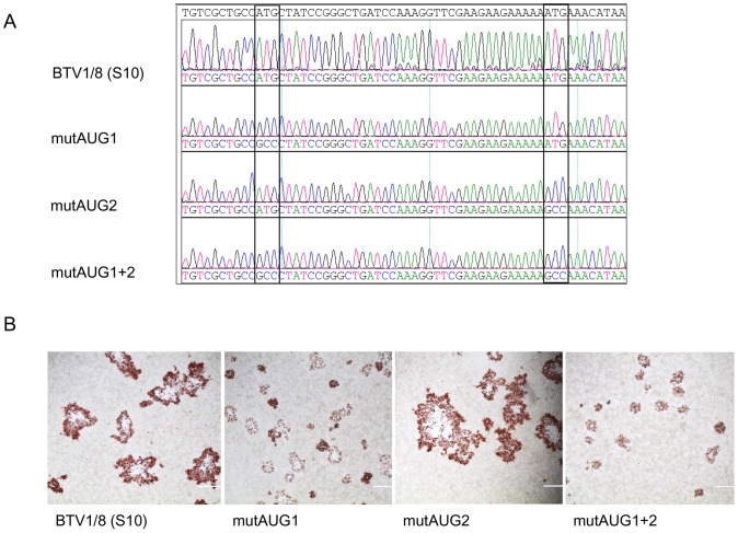
Figure 3. Sequence analysis and plaque phenotype of AUG mutant viruses.
(A) Sequence analysis of Seg-10 amplicons from AUG mutant viruses shown in DNASTAR Lasergene Seqman assembly software. The mutated codons are indicated in rectangles (B) Plaque morphology of BSR cells infected with wtBTV1/8(S10), mutAUG1, mutAUG2 and mutAUG1+2 virus grown under 1% methylcellulose overlay medium are shown. At 2 dpi cells were fixed and immunostained with anti-VP7 Mab.

