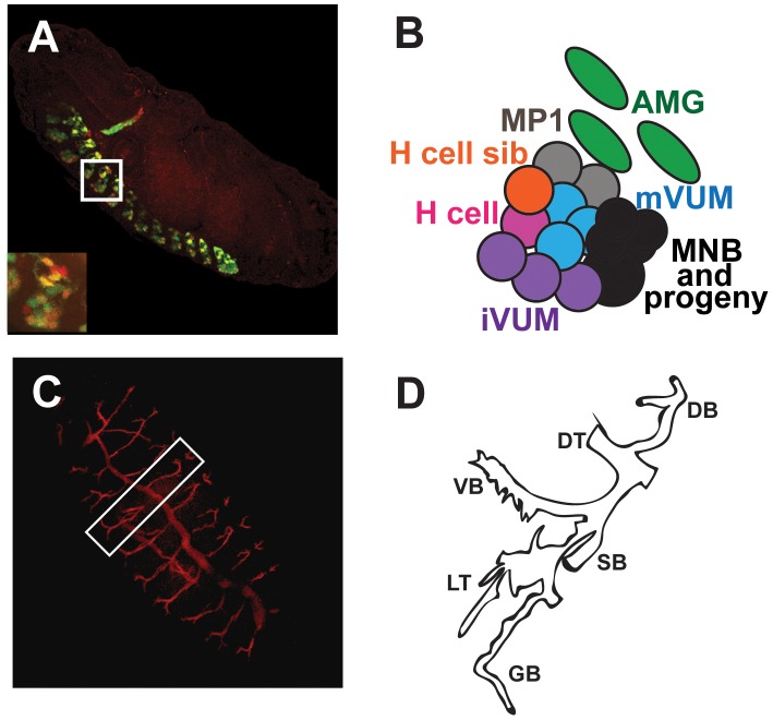
Figure 1. Relative locations of the CNS midline and trachea within the late Drosophila embyo.
(A) The midline cellular pattern is segmentally repeated throughout the ventral nerve cord at embryonic stage 16. (B) Each segment consists of six neural subtypes and three surviving midline glia whose relative locations within a typical thoracic segment (white box and inset in A) are shown. The midline subtypes include: the MP1 neurons (gray), the H cell (pink), the H cell sib (orange), the ventral unpaired interneurons (iVUMs; purple), the ventral unpaired motorneurons (mVUMs; blue), median neuroblast (MNB) and its progeny (black) and the anterior midline glia (AMG; green); adapted from [24], [108]. (C) By the end of embryogenesis, the trachea form an extensive network that mediates gas exchange throughout the organism. (D) Each tracheal metamere consists of the major dorsal trunk (DT), a dorsal branch (DB), and the visceral (VB), spiracular (SB) and ganglionic (GB) branches and lateral trunk (LT) on the ventral side; adapted from [71]. Lateral views of whole mount embryos stained with anti-GFP (green), anti-sim (red; A) antibodies or monoclonal antibody 2A12 (red; C) and analyzed by confocal microscopy are shown. (A) The embryo contains a reporter gene that expresses GFP in all midline cells.

