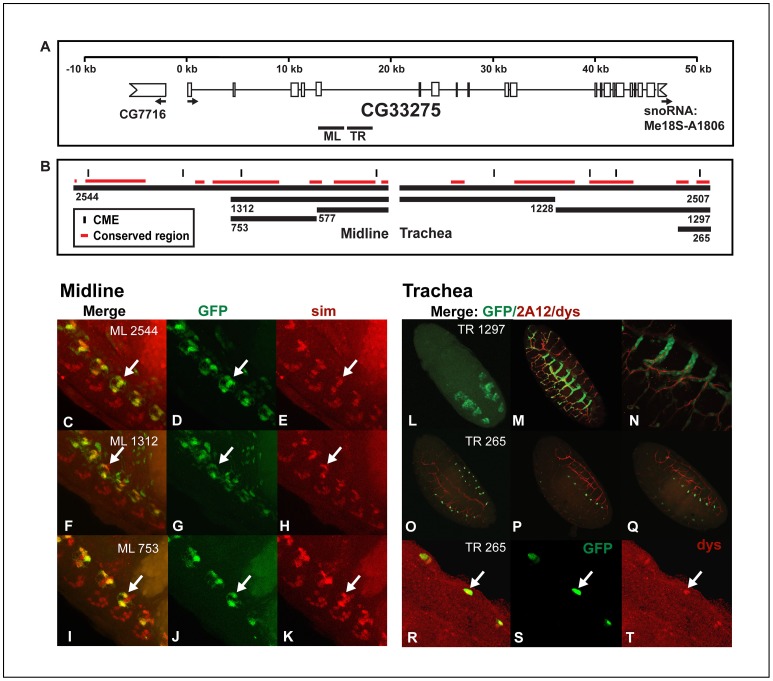
Figure 2. CG33275 contains a midline enhancer that is separable and distinct from a nearby tracheal enhancer.
(A) Locations of regions within the fifth intron of CG33275 used to generate the reporter constructs are shown. A scale is indicated on top and the thick lines represent the regions analyzed in (B–T). White boxes represent exons and thin lines represent introns. The indented boxes indicate flanking genes, CG7716 and snoRNA, and arrows indicate the start and direction of transcription. (B) Fragments used to generate reporter constructs are shown. The size of each fragment is indicated below it, vertical lines indicate locations of CMEs and red lines represent sequence blocks conserved in at least 11 Drosophila species. (C–T) Whole mount embryos were double-stained with anti-GFP (green: D, G, J, L and S), anti-sim (red; E, H, K), anti-dys (red; T) antibodies and monoclonal antibody 2A12 (red; M–Q) and analyzed by confocal microscopy. The overlap in expression is shown in yellow in the merge images (C, F, I, M–Q and R). Reporters (C–E) CG33275 ML2544:GFP, (F–H) CG33275 ML1312:GFP and (I–K) CG33275 ML753:GFP drove expression in midline glia. Midline glia can be identified by the overlap in expression of GFP and sim (arrows C–K) and are located on the dorsal side of the nerve chord. Midline neurons are located on the ventral side of the nerve chord and labeled by sim, but do not express CG33275 or any of the CG33275 reporter genes. (L–T) Monoclonal antibody 2A12 labels the tracheal lumen and the anti-dys antibody labels tracheal fusion cells. CG33275 TR1297:GFP is expressed in all trachea, beginning in the tracheal pits at stage 12 (L) and extending to late embryogenesis (M–N), while CG33275 TR265:GFP is expressed only in fusion cells (O–T), indicated by co-localization of GFP and Dys (arrows R–T). Lateral views of stage 16 transgenic embryos are shown; anterior is in the top, left hand corner and ventral is on the left, except (L), which is a dorsal view of a stage 12 embryo.

