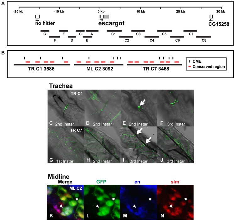
Figure 3. An esg genomic region contains a midline enhancer that is separable and distinct from two esg tracheal enhancers.
(A) Genomic regions surrounding esg used to generate the reporter constructs and (B) the esg enhancers are shown as above. Gray boxes represent 5′ and 3′ untranslated regions. (C–J) Live larvae were analyzed by confocal and differential contrast microscopy and ventral views of the (C–F) esg TR C1:GFP and (G–J) esg TR C7:GFP reporters are shown. In larvae, the esg TR C1:GFP reporter is expressed sporadically in fusion cells (arrow in E) and the esg TR C7:GFP reporter is expressed in all tracheal branches and sporadically in the dorsal trunks (G–J), but consistently in fusion cells (arrows I). (K–N) Whole mount esg ML C2:GFP reporter embryos were stained with an anti-GFP antibody (green: L), engrailed monoclonal antibody (blue; M) and anti-sim antibody (red; N) and analyzed by confocal microscopy. The overlap in expression is shown in the merge image (K). Anterior midline glia express GFP and sim and are located dorsally within the nerve chord. Posterior midline glia that normally undergo cell death during this time can still be visualized with GFP (three cells surrounding star in N), but not sim or engrailed. The MNB and its progeny express sim, engrailed and GFP and are located ventrally within the nerve chord (arrowheads in K–N). Lateral view of a stage 16 transgenic embryo is shown; anterior is in the top, left hand corner and ventral is on the left.

