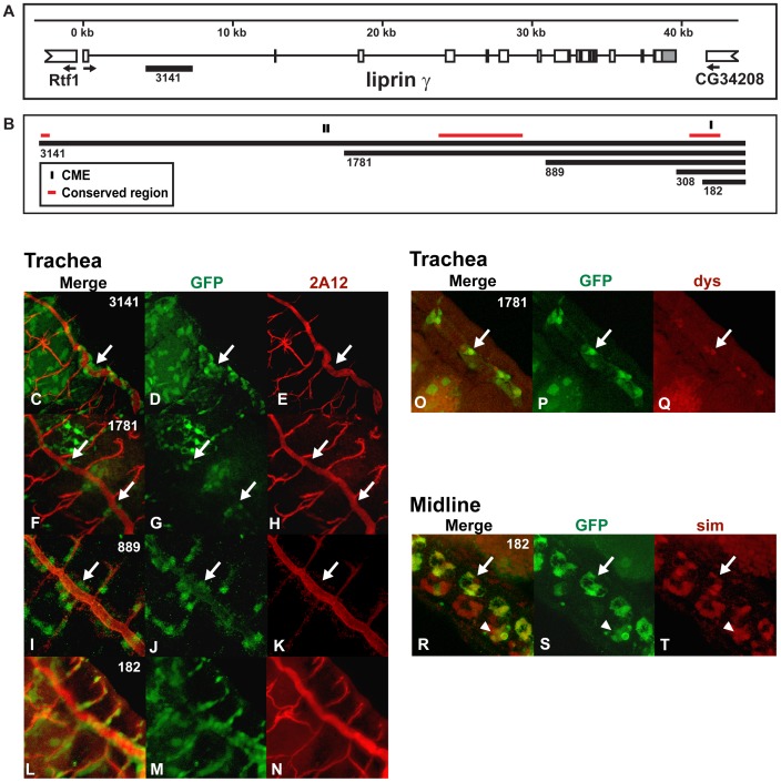
Figure 4. Liprin γ contains a conserved enhancer sufficient to drive expression in both midline glia and trachea.
(A) The genomic regions within the first intron of liprin γ used to generate the reporter constructs and (B) the liprin γ enhancers are shown as in Fig. 1 and previously reported [59]. (C–T) Whole mount embryos were double-stained with an anti-GFP antibody (green: D, G, J, M, P and S) and monoclonal antibody 2A12 (red; E, H, K and N), anti-dys (red; Q) or anti-sim (red; T) and analyzed by confocal microscopy. The overlap in expression is shown in yellow in the merge column (C, F, I, L, O and R). Even though the liprin γ 3141:GFP and liprin γ 889:GFP reporters are expressed in the dorsal trunk (arrows C–E and I–K) and dorsal and ventral branches, liprin γ 1781:GFP is restricted to mostly fusion cells of the dorsal trunk (arrows F–H and O–Q). The liprin γ 182:GFP reporter is expressed in all tracheal cells (L–N), midline glia (arrows R–T) and a few midline neurons in certain segments (arrowheads R–T). Lateral views of stage 16 transgenic embryos are shown; anterior is in the top, left hand corner and ventral is on the left.

