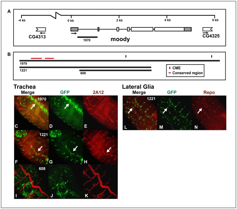
Figure 7. The moody cis-regulatory region contains a tracheal enhancer that overlaps with a lateral CNS glial enhancer.
(A) The genomic regions surrounding and within moody used to generate the reporter constructs and (B) the moody enhancer is shown. (C–N) Whole mount embryos were double-stained with an anti-GFP antibody (green: D, G, J, and M) and monoclonal antibody 2A12 (red; E, H and K) or anti-repo monoclonal antibody (red; N) and analyzed by confocal microscopy. The overlap in expression is shown in yellow in the merge columns (C, F, I and L). The expression patterns of the (C–E) moody1970:GFP, (F–H and L–N) moody1221:GFP and (I–K) moody608:GFP reporters are shown. Both the moody1970:GFP (not shown) and moody1221:GFP (L–N) reporters also drive expression in lateral glia as indicated by co-localization with repo (arrows). Additionally, moody 1221:GFP is expressed in the dorsal trunk (arrows F–H), while moody1970:GFP is expressed in the dorsal vessel (arrows C and D) and lightly in the dorsal trunk. (C–H) Dorsolateral, (I–K) lateral or (L–N) ventral views of stage 16 transgenic embryos are shown; anterior is in the top, left hand corner.

