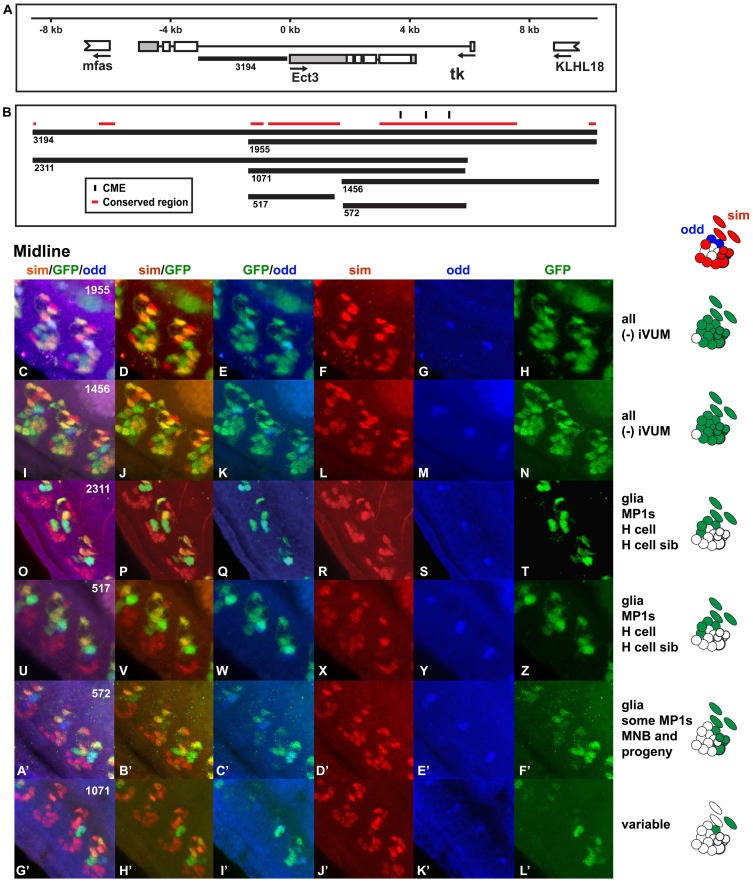
Figure 8. The Ect3 cis-regulatory region contains a midline enhancer sensitive to context.
(A) The genomic region upstream of Ect3 (and within the first intron of Tk) used to generate the reporter constructs and (B) the Ect3 enhancers are shown. (C–L’) Whole mount embryos were stained with anti-sim (red; F, L, R, X, D’ and J’), anti-odd (blue; G, M, S, Y, E’ and K’) and anti-GFP (green: H, N, T, Z, F’ and L’) antibodies and analyzed by confocal microscopy. The overlap in expression is shown in the merge columns: all three antibodies (C, I, O, U, A’ and G’), sim and GFP (D, J, P, V, B’ and H’) and GFP and odd (E, K, Q, W, C’ and I’). Odd is expressed only in MP1 midline neurons. Both Ect3 1955:GFP (C–H) and Ect3 1456:GFP (I–N) drive expression in the all midline cells, with the exception of a single iVUM. Ect3 2311:GFP (O–T) and Ect3 517:GFP (U–Z) are restricted to some midline glia, MP1 neurons, the H cell and H cell sib. Ect3 572:GFP (A’–F’) is expressed in midline glia, some MP1 neurons and some of the MNB and its progeny. Finally, Ect3 1071:GFP (G’–L’) drives only spotty midline expression. Lateral views of stage 16 transgenic embryos are shown; anterior is in the top, left hand corner and ventral is on the left. The midline expression pattern of each reporter is shown schematically on the right.

