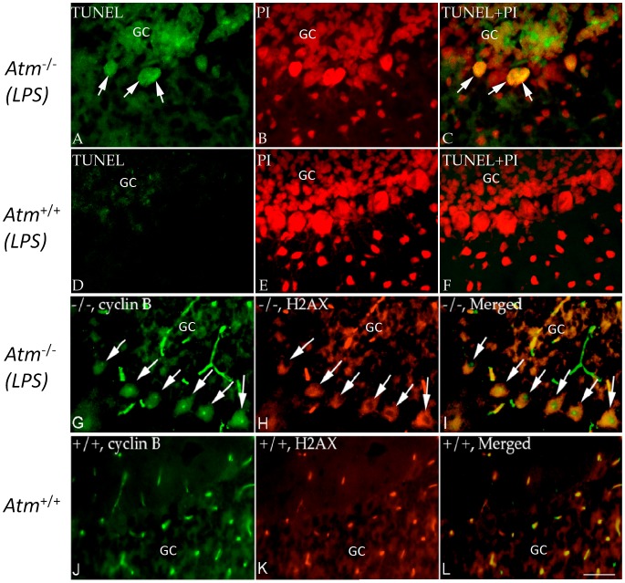
Figure 6. Appearance of cell death markers in Atm−/− mice after LPS treatment.
The representative pictures from each group (n = 3–4, repeated 3 times) were shown. (A–F) TUNEL staining was found in a small number of Atm−/− Purkinje cells (A–C), but not in wild type controls (D–F) after 3 days of LPS treatment. TUNEL preparations were counterstained with PI. After 4 weeks of LPS treatment, many Atm−/− Purkinje cells expressed cyclin B (green, G–I) and those that did also co-localized with γ-H2AX in mice (red). In wild type controls (J–L) nuclear cyclin B and γ-H2AX staining were rare. Scale bar = 50 µm.

