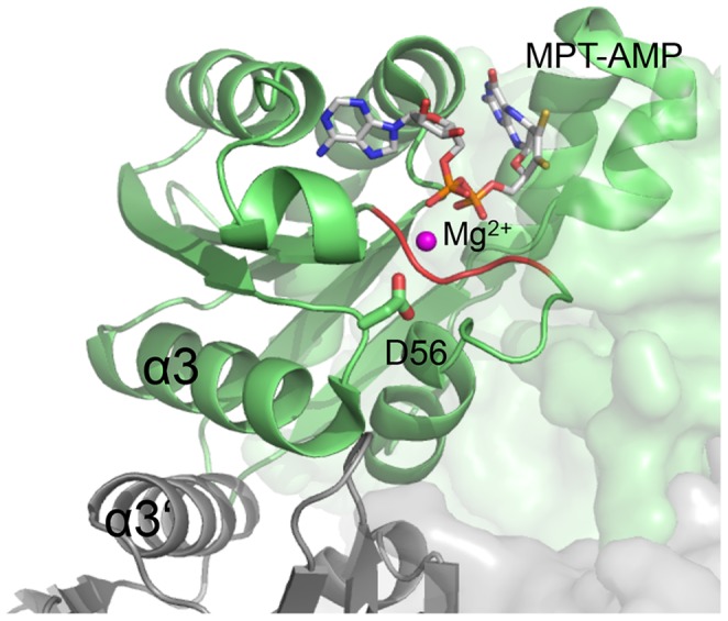
Figure 7. Active site of PfuMoaB-WT.

Two PfuMoaB subunits at the hexamerization interface are shown as ribbon in green and grey, respectively. The conserved Asp56 residue coordinating Mg2+ (pink) is shown in sticks. MPT-AMP in the active site is derived from a superimposition with the structure of the PfuMoaB homologue A. thaliana Cnx1G (1UUY). The Mg2+-ion derived from a superimposition with the homologues sub-domain 3 of E. coli MoeA (1FC5) [6], [63].
