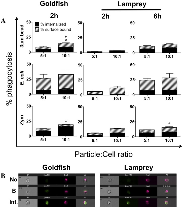
Figure 1. Phagocytosis of different target particles by goldfish and lamprey primary leukocytes.
(A) Goldfish primary kidney leukocytes (PKL) or lamprey primary typhlosole leukocytes (PTL) were incubated with 3 µm YG latex beads, E. coli DH5α-GFP, or zymosan-FITC at the indicated concentrations for the specified times. Cells were then fixed and phagocytosis was quantified by flow cytometry. Grey bars represent percent internalized. Hatched white bars represent percent surface bound. For all n = 4, over 2 examined over a minimum of two independent experiments. * p<0.05 for % internalized,+p<0.05 for % surface bound- between 10∶1 and 5∶1 particle to cell ratios in each graph. (B) Representative images of no internalization (No), surface bound (B), and internalized beads (Int.) from ImageStream MkII flow cytometer (Amnis).

