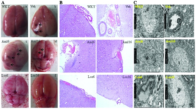
Figure 2.
Changes in the brain structure of Wistar Kyoto rats (WKY), stroke-prone spontaneously hypertensive rats (SHRSP)-Veh and the four drug treatment SHRSP groups at the age of 40 weeks. (A) A global view of brains dissected from rats of each group investigated. Arrows point to the bleeding spots. Note that no obvious bleeding spots were observed in the brains from the SHRSP-Los6 and -Los16 groups. (B) Hematoxylin and eosin (H&E) staining shows bleeding and/or disorganized brain cells in the SHRSP-Veh, SHRSP-Los6, and two amlodipine treatment groups, but not the SHRSP-Los16 group. (C) Ultrastructural analysis of brain tissues from the six groups by transmission electron microscopy shows swollen mitochondria and disrupted cristae in SHRSP-Veh. Similar pathological changes are present in the SHRSP-Aml6, SHRSP-Aml16 and SHRSP-Los6 groups, but not in the SHRSP-Los16 and WKY groups. (A–C) Representative data are shown.

