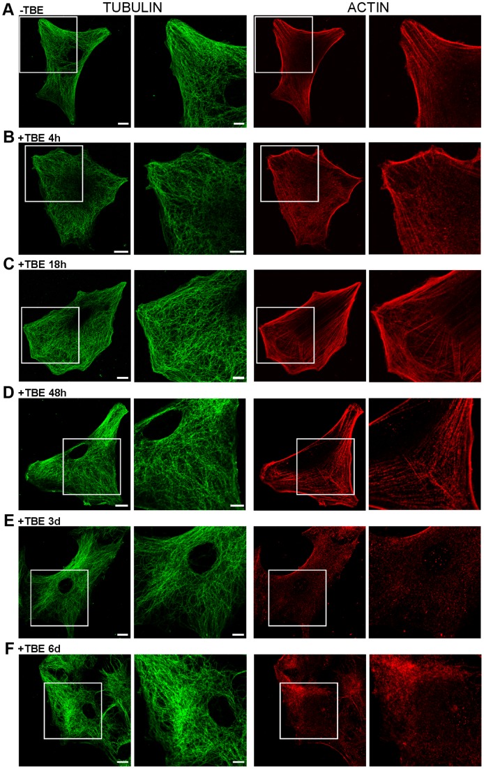
Figure 6. TBEV affected cytoskeleton morphology, but not cell shape nor cell viability.
A–F, Immunocytochemical labellings of TUBULIN (anti-α-Tubulin 1∶100) and ACTIN cytoskeleton (anti-β-Actin 1∶200). Squared area is 2× enlarged in adjacent columns. Substantial rearrangement of actin cytoskeleton was observed after 3 and 6 days p.i. (E,F). Cell shape remained preserved at all times p.i. Bars: 10 µm (whole cell), 5 µm (enlarged panels).

