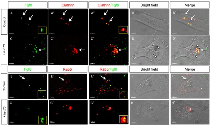
Figure 3. Analysis of Fgf8 internalization Clathrin coated vesicles and early endosomes in fish fibroblasts.
Confocal analysis of Fgf8-GFP co-localization with indicated early endocytic markers in zebrafish PAC2 cells. Columns 1–3 show confocal images and columns 4–5 shows bright field images merged with the confocal image. Co-localization of Fgf8 with Clathrin (A–D’) and with Rab5 (E–H’) was investigated. Furthermore, insets show the increasing size of Rab5 positive early endosomes upon Hsc70 overexpression (E and G).

