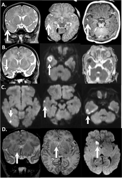
Figure 1.
Four HLHS patients found to have stroke (white arrows) on pre-operative brain MRI: Images read left to right. A: coronal and axial T2 demonstrating hyperintense signal in the right temporal lobe, involving the cortex, hemorrhage seen as a hypointensity on T2 and hyperintensity on axial T1 (3rd image on right). B: Similar to A, Coronal T2 (left) shows hemorrhage inside stroke, Diffusion weighted imaging (DWI) showing restriction of water diffusion (center) with Apparent diffusion coefficient (ADC) confirmation (right). C: DWI demonstrating multifocal infarcts in the right temporal lobe. D: Head of the caudate nucleus stroke seen on coronal and axial T2 (left and center) with confirmation of restriction of water diffusion on axial DWI.

