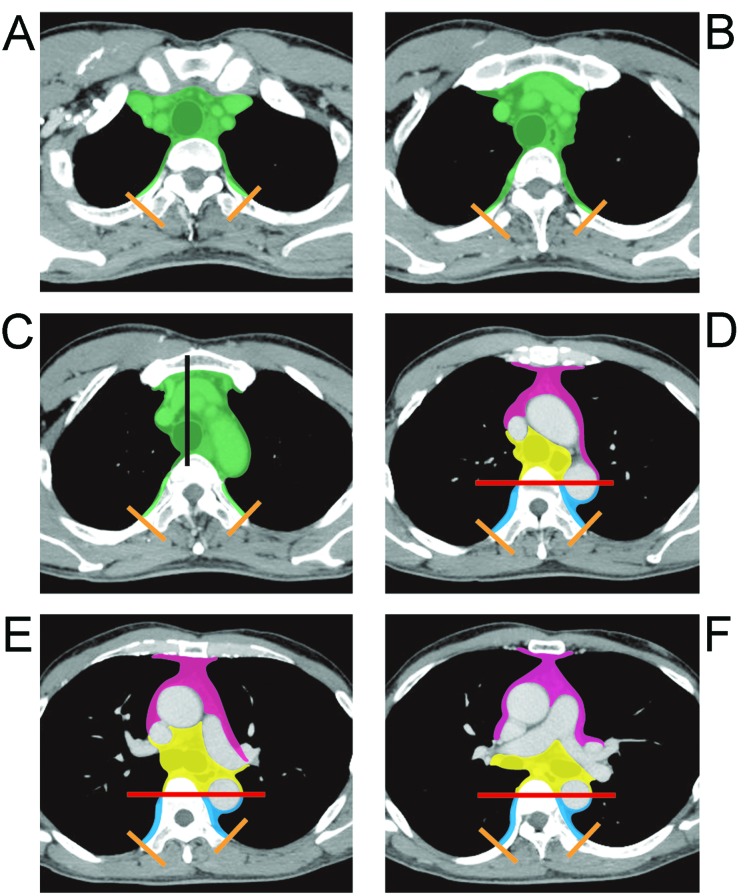
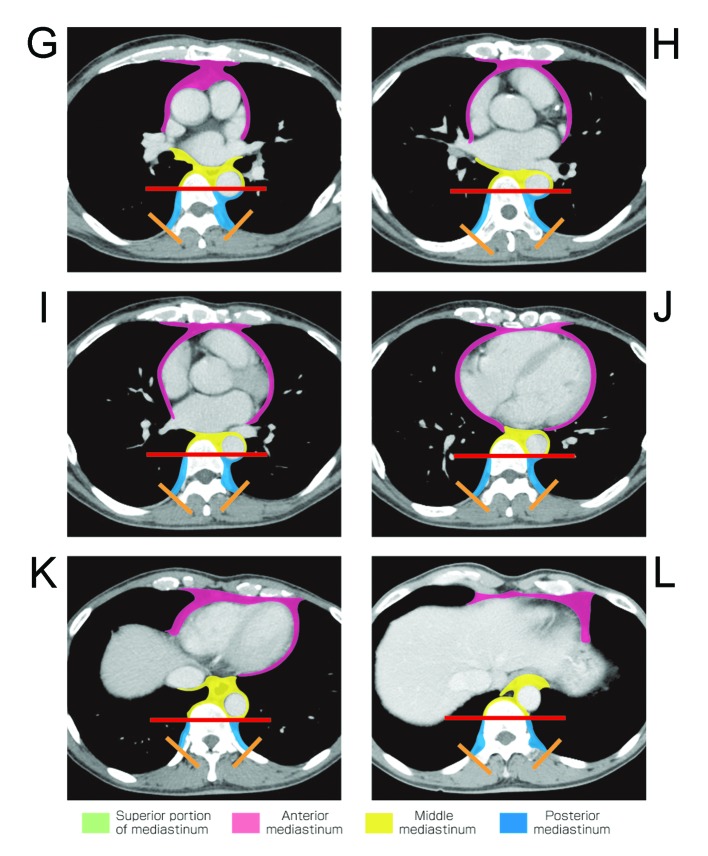
Figure 2.
Contrast-enhanced CT images represent new proposal of mediastinal compartment classification according to the General Rules for Study of Mediastinal Tumors of the JART. (A) Thoracic inlet, (B) upper rim of clavicle, (C) sterno-clavicular joint, (D) left brachiocephalic vein across TML, (E) aortic arch, (F) tracheal carina, (G) right main pulmonary artery, (H) pulmonary trunk, (I) left atrium, (J) tricuspid valve, (K) hepatic dome of diaphragm, (L) middle of 12th thoracic vertebral body. Abbreviations: TML, tracheal mid-line; M-PBL, middle-posterior boundaryline (see Materials and methods).


