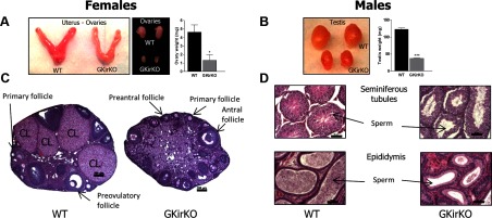
Figure 5.

Gross gonadal anatomy and gonadal histology of GKirKO mice. A, Small ovaries (size and weight) and uterus found in GKirKO mice compared with WT. B, Smaller size and weight of the testes in GKirKO male mice compared with WT mice. C, Representative sections from a WT ovary (left image) showing follicles at all stages of development, including primary, preantral, antral, preovulatory follicles as well as corpora lutea (CL); GKirKO mice (right image), in contrast, do not contain follicles past the antral stage and have no corpora lutea formation. Scale bars correspond to 100 μm. The most representative microphotographs were chosen. D, Representative sections of testes. Seminiferous tubules from a WT mouse (left, top image) show all stages of spermatogenesis with numerous sperm. In contrast, seminiferous tubules from GKirKO mice (right, top image) lack spermatogenesis. Representative sections of the epididymis: the lumen from a WT mouse is filled with sperm (left, bottom image), whereas that of GKirKO mice have fewer sperm (right, bottom image). Scale bars represent 50 μm.
