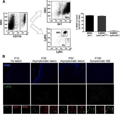
Figure 1. MDSCs are present in medulloblastoma tumors from Smo* mice.

(A) Flow cytometry analysis of immune cell suspensions prepared from adult Smo* mice medulloblastoma tumors (n=9) using CD11b, Gr1, Ly6G, and Ly6C markers. Cells were first gated on the CD11b+ population and then analyzed for Gr1 or Ly6C/Ly6G expression. Representative FACS plots (left) and average quantification (right) are shown. Graph shows percent of cells within the CD11b+ population. SSC, Side-scatter. (B) Immunofluorescence analysis of CD11b+Ly6G+ cell presence in cerebellar lesions of Smo* mice collected at different stages: Postnatal Day (P) 10 (no lesions observed, preserving normal cerebellar structures), P28 (cell-dense, ectopic lesions are observed), P50, and P180 (symptomatic, fully developed tumor is present). CD11b, red; Ly6G, green; nuclei (DAPI), blue. One experiment from three is shown. Original scale bar, 100 μm.
