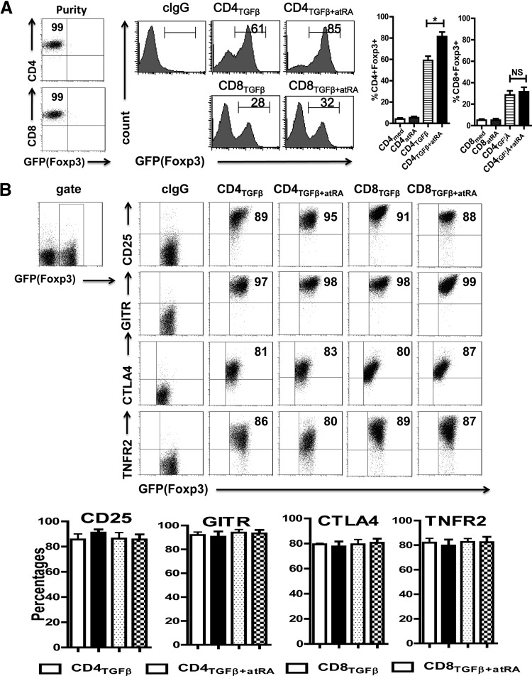
Figure 1. ATRA increased the percentages of Foxp3 expression on TGF-β-primed CD4+, but not on CD8+ cells.
(A) CD8+CD62L+CD25−Foxp3−(GFP−) and CD4+CD62L+CD25−Foxp3−(GFP−) cells isolated from C57BL/6 Foxp3gfp reporter mice were stimulated with immobilized anti-CD3 (1 μg/ml), soluble anti-CD28 (1 μg/ml), IL-2 (100 U/ml), or TGF-β (2 ng/ml), with (CD4TGFβ+ATRA or CD8TGFβ+ATRA) or without ATRA (50 nM) (CD4TGFβ or CD8TGFβ) for 3 days. Foxp3 (GFP) expression was examined by flow cytometry. Left: typical FACS histograms. Right: summary of data showing the frequency of Foxp3+ cells from TGF-β-primed CD4+ or CD8+ cells. *P < 0.05, NS. (B) The expression levels of regulatory T-cell associated markers including CD25, GITR, CTLA-4, and TNFR2 on CD4TGFβ, CD8TGFβ, CD4TGFβ+ATRA, or CD8TGFβ+ATRA cells were analyzed by flow cytometry. The graph data indicate the mean ± sem of 3 separate experiments showing the frequency of the indicated markers gated on the CD4 or CD8 cell populations.

