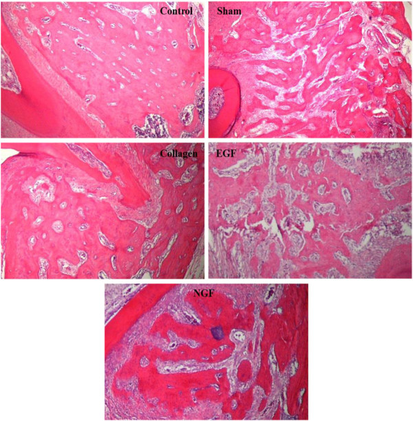
Figure 10.

Representative photomicrographs demonstrating inter-radicular alveolar bone architecture of the maxillary first molars. Photomicrographs (×40 H & E) of the inter-radicular alveolar bone of the upper right first molar from the experimental groups (sham, collagen, EGF and NGF) and a representative photomicrograph of the upper left first molar inter-radicular alveolar bone (control). The control photomicrograph shows the normal architecture of the alveolar process with thick bone trabecula and small medullary spaces. Photomicrographs of the experimental groups (sham, collagen, EGF and NGF) show a fragmented alveolar process consisting of thin bone trabecular and wide medullary spaces.
